| dc.contributor.author | Bakke, Kine Mari | |
| dc.contributor.author | Meltzer, Sebastian | |
| dc.contributor.author | Grøvik, Endre | |
| dc.contributor.author | Negård, Anne | |
| dc.contributor.author | Holmedal, Stein Harald | |
| dc.contributor.author | Gjesdal, Kjell-Inge | |
| dc.contributor.author | Bjørnerud, Atle | |
| dc.contributor.author | Ree, Anne Hansen | |
| dc.contributor.author | Redalen, Kathrine | |
| dc.date.accessioned | 2021-08-05T13:06:58Z | |
| dc.date.available | 2021-08-05T13:06:58Z | |
| dc.date.created | 2020-11-25T22:33:08Z | |
| dc.date.issued | 2020 | |
| dc.identifier.citation | Bakke, K. M., Meltzer, S., Grøvik, E., Negård, A., Holmedal, S. H., Gjesdal, K.-I., Bjørnerud, A., Ree, A. H. & Redalen, K. R. (2020). Sex Differences and Tumor Blood Flow from Dynamic Susceptibility Contrast MRI Are Associated with Treatment Response after Chemoradiation and Long-term Survival in Rectal Cancer. Radiology, 297(2), 352-360. | en_US |
| dc.identifier.issn | 0033-8419 | |
| dc.identifier.uri | https://hdl.handle.net/11250/2766551 | |
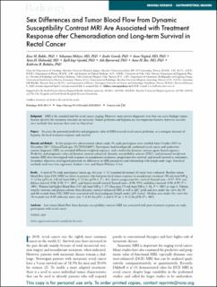
| dc.description.abstract | Sex differences, tumor vascularity, and blood flow seen on multiecho dynamic contrast–based MRI scans enabled prediction of treatment response and overall survival in patients with rectal cancer.
Background: MRI is the standard tool for rectal cancer staging. However, more precise diagnostic tests that can assess biologic tumor features decisive for treatment outcome are necessary. Tumor perfusion and hypoxia are two important features; however, no reference methods that measure these exist in clinical use.
Purpose: To assess the potential predictive and prognostic value of MRI-assessed rectal cancer perfusion, as a surrogate measure of hypoxia, for local treatment response and survival.
Materials and Methods: In this prospective observational cohort study, 94 study participants were enrolled from October 2013 to December 2017 (ClinicalTrials.gov: NCT01816607). Participants had histologically confirmed rectal cancer and underwent routine diagnostic MRI, an extended diffusion-weighted sequence, and a multiecho dynamic contrast agent–based sequence. Predictive and prognostic values of dynamic contrast-enhanced, dynamic susceptibility contrast (DSC), and intravoxel incoherent motion MRI were investigated with response to neoadjuvant treatment, progression-free survival, and overall survival as end points. Secondary objectives investigated potential sex differences in MRI parameters and relationship with lymph node stage. Statistical methods used were Cox regression, Student t test, and Mann-Whitney U test.
Results: A total of 94 study participants (mean age, 64 years ± 11 [standard deviation]; 61 men) were evaluated. Baseline tumor blood flow from DSC MRI was lower in patients who had poor local tumor response to neoadjuvant treatment (96 mL/min/100 g ± 33 for ypT2–4, 120 mL/min/100 g ± 21 for ypT0–1; P = .01), shorter progression-free survival (hazard ratio = 0.97; 95% confidence interval: 0.96, 0.98; P < .001), and shorter overall survival (hazard ratio = 0.98; 95% confidence interval: 0.98, 0.99; P < .001). Women had higher blood flow (125 mL/min/100 g ± 27) than men (74 mL/min/100 g ± 26, P < .001) at stage 4. Volume transfer constant and plasma volume from dynamic contrast-enhanced MRI as well as ΔR2* peak and area under the curve for 30 and 60 seconds from DSC MRI were associated with local malignant lymph nodes (pN status). Median area under the curve for 30 seconds was 0.09 arbitrary units (au) ± 0.03 for pN1–2 and 0.19 au ± 0.12 for pN0 (P = .001).
Conclusion: Low tumor blood flow from dynamic susceptibility contrast MRI was associated with poor treatment response in study participants with rectal cancer. | en_US |
| dc.language.iso | eng | en_US |
| dc.subject | Magnetisk resonans tomografi, MRI | en_US |
| dc.subject | Magnetic Resonance Imaging, MRI | en_US |
| dc.subject | Kreftdiagnostikk | en_US |
| dc.subject | Cancer diagnostics | en_US |
| dc.title | Sex differences and tumor blood flow from dynamic susceptibility contrast MRI are associated with treatment response after chemoradiation and long-term survival in rectal cancer | en_US |
| dc.type | Peer reviewed | en_US |
| dc.type | Journal article | en_US |
| dc.description.version | publishedVersion | en_US |
| dc.rights.holder | © RSNA, 2020. | en_US |
| dc.subject.nsi | VDP::Medisinsk teknologi: 620 | en_US |
| dc.subject.nsi | VDP::Medical technology: 620 | en_US |
| dc.subject.nsi | VDP::Medisinsk teknologi: 620 | en_US |
| dc.subject.nsi | VDP::Medical technology: 620 | en_US |
| dc.source.pagenumber | 352-360 | en_US |
| dc.source.volume | 297 | en_US |
| dc.source.journal | Radiology | en_US |
| dc.source.issue | 2 | en_US |
| dc.identifier.doi | https://doi.org/10.1148/RADIOL.2020200287 | |
| dc.identifier.cristin | 1852539 | |
| dc.relation.project | Akershus universitetssykehus HF: 267940, 268938 | en_US |
| dc.relation.project | Helse Sør-Øst RHF: 2014012, 2015048, 2016050 | en_US |
| dc.relation.project | Kreftforeningen: 198116-2018 | en_US |
| cristin.ispublished | true | |
| cristin.fulltext | postprint | |
| cristin.qualitycode | 2 | |