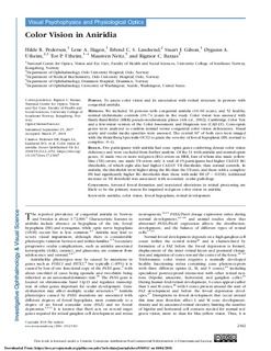
Color Vision in Aniridia
| dc.contributor.author | Pedersen, Hilde Røgeberg | |
| dc.contributor.author | Hagen, Lene Aarvelta | |
| dc.contributor.author | Landsend, Erlend Christoffer Sommer | |
| dc.contributor.author | Gilson, Stuart | |
| dc.contributor.author | Utheim, Øygunn Aass | |
| dc.contributor.author | Utheim, Tor Paaske | |
| dc.contributor.author | Neitz, Maureen | |
| dc.contributor.author | Baraas, Rigmor C. | |
| dc.date.accessioned | 2018-10-05T06:39:15Z | |
| dc.date.available | 2018-10-05T06:39:15Z | |
| dc.date.created | 2018-10-02T11:06:33Z | |
| dc.date.issued | 2018 | |
| dc.identifier.citation | Investigative Ophthalmology and Visual Science. 2018, 59 (5), 2142-2152. | nb_NO |
| dc.identifier.issn | 0146-0404 | |
| dc.identifier.uri | http://hdl.handle.net/11250/2566543 | |
| dc.description | This work is licensed under a Creative Commons Attribution-NonCommercial-NoDerivatives 4.0 International License. | nb_NO |
| dc.description.abstract | Purpose: To assess color vision and its association with retinal structure in persons with congenital aniridia. Methods: We included 36 persons with congenital aniridia (10–66 years), and 52 healthy, normal trichromatic controls (10–74 years) in the study. Color vision was assessed with Hardy-Rand-Rittler (HRR) pseudo-isochromatic plates (4th ed., 2002); Cambridge Color Test and a low-vision version of the Color Assessment and Diagnosis test (CAD-LV). Cone-opsin genes were analyzed to confirm normal versus congenital color vision deficiencies. Visual acuity and ocular media opacities were assessed. The central 30° of both eyes were imaged with the Heidelberg Spectralis OCT2 to grade the severity of foveal hypoplasia (FH, normal to complete: 0–4). Results: Five participants with aniridia had cone opsin genes conferring deutan color vision deficiency and were excluded from further analysis. Of the 31 with aniridia and normal opsin genes, 11 made two or more red-green (RG) errors on HRR, four of whom also made yellow-blue (YB) errors; one made YB errors only. A total of 19 participants had higher CAD-LV RG thresholds, of which eight also had higher CAD-LV YB thresholds, than normal controls. In aniridia, the thresholds were higher along the RG than the YB axis, and those with a complete FH had significantly higher RG thresholds than those with mild FH (P = 0.038). Additional increase in YB threshold was associated with secondary ocular pathology. Conclusions: Arrested foveal formation and associated alterations in retinal processing are likely to be the primary reason for impaired red-green color vision in aniridia. | nb_NO |
| dc.language.iso | eng | nb_NO |
| dc.relation.uri | https://iovs.arvojournals.org/article.aspx?articleid=2679803&resultClick=1 | |
| dc.rights | Attribution-NonCommercial-NoDerivatives 4.0 Internasjonal | * |
| dc.rights.uri | http://creativecommons.org/licenses/by-nc-nd/4.0/deed.no | * |
| dc.title | Color Vision in Aniridia | nb_NO |
| dc.title.alternative | Color Vision in Aniridia | nb_NO |
| dc.type | Journal article | nb_NO |
| dc.type | Peer reviewed | nb_NO |
| dc.description.version | publishedVersion | nb_NO |
| dc.rights.holder | (c) The Authors | nb_NO |
| dc.source.pagenumber | 2142-2152 | nb_NO |
| dc.source.volume | 59 | nb_NO |
| dc.source.journal | Investigative Ophthalmology and Visual Science | nb_NO |
| dc.source.issue | 5 | nb_NO |
| dc.identifier.doi | 10.1167/iovs.17-23047 | |
| dc.identifier.cristin | 1617092 | |
| cristin.unitcode | 222,56,2,0 | |
| cristin.unitname | Institutt for optometri, radiografi og lysdesign | |
| cristin.ispublished | true | |
| cristin.fulltext | original | |
| cristin.qualitycode | 2 |
Tilhørende fil(er)
Denne innførselen finnes i følgende samling(er)

Med mindre annet er angitt, så er denne innførselen lisensiert som Attribution-NonCommercial-NoDerivatives 4.0 Internasjonal