| dc.contributor.author | Pedersen, Hilde Røgeberg | |
| dc.date.accessioned | 2020-05-04T06:38:23Z | |
| dc.date.available | 2020-05-04T06:38:23Z | |
| dc.date.issued | 2020-05-12 | |
| dc.identifier.isbn | 978-82-7206-553-8 | |
| dc.identifier.issn | 2535-5252 | |
| dc.identifier.uri | https://hdl.handle.net/11250/2653117 | |
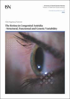
| dc.description.abstract | Aniridia is a rare, congenital eye disorder most commonly caused by a mutation in the PAX6 gene, which affects eye development and leads to a range of ocular anomalies, including iris- and foveal hypoplasia and vision impairment. However, the phenotypes vary considerably between individuals. Research investigating the retina in aniridia remains limited. The main purpose of this thesis is therefore to gain more in-depth knowledge about variation in genotype and retinal phenotype in persons with aniridia.
The thesis includes three cross-sectional studies that characterize macular structure and foveal development, their importance to visual performance, and genotype-phenotype correlations. Data from genetic analysis and retinal imaging were combined with clinical and psychophysical measures of high-contrast visual acuity and colour vision.
High-resolution retinal imaging shows that persons with aniridia have varying foveal hypoplasia grades (paper I), reduced cone photoreceptor density and mosaic regularity (paper II) and decreased thicknesses and morphology of the retinal layers (paper III), relative to normal healthy controls. High-contrast visual acuity and colour discrimination thresholds not only varied greatly between individuals, but also within families carrying the same genetic mutation, and were associated with grade of foveal hypoplasia and thickness of the outer retinal layers. Despite the large variation in phenotype, analysis of genotype-phenotype correlations indicate that the retinal phenotype is associated with the position and extent of the mutation, within non-coding, coding or flanking regulatory regions of the PAX6 gene.
This knowledge is of great significance in the clinical management of persons with congenital aniridia to understand limits and potential related to visual function, to determine when an intervention is advisable, and for presenting well-founded individual alternatives of facilitation, rehabilitation or treatment options. | en_US |
| dc.language.iso | eng | en_US |
| dc.publisher | University of South-Eastern Norway | en_US |
| dc.relation.ispartofseries | Doctoral dissertations at the University of South-Eastern Norway;67 | |
| dc.relation.haspart | Paper I: Pedersen, H.R., Hagen, L.A., Landsend, E.C.S., Gilson, S.J., Utheim, Ø.A., Utheim, T.P., Neitz M. & Baraas, R.C.: Color Vision in Aniridia. Investigative Ophthalmology & Visual Science, 59(5), 2142-2152, (2018). https://doi.org/10.1167/iovs.17-23047 | en_US |
| dc.relation.haspart | Paper II: Pedersen, H.R., Neitz, M., Gilson, S.J., Landsend, E.C.S., Utheim, Ø.A., Utheim, T.P., & Baraas, R.C.: The cone photoreceptor mosaic in aniridia: within-family phenotype-genotype discordance. Opthalmology Retina, 3(6), 523-534, (2019). https://doi.org/10.1016/j.oret.2019.01.020 | en_US |
| dc.relation.haspart | Paper III: Pedersen, H.R., Baraas, R.C., Landsend, E.C.S., Utheim, Ø.A., Utheim, T.P., Gilson, S.J. & Neitz M.: PAX6 Genotypic and Retinal Phenotypic Characterization in Congenital Aniridia. Accepted for publication in Investigative Ophthalmology & Visual Science. | en_US |
| dc.rights | Navngivelse-Ikkekommersiell-DelPåSammeVilkår 4.0 Internasjonal | * |
| dc.rights.uri | http://creativecommons.org/licenses/by-nc-sa/4.0/deed.no | * |
| dc.subject | aniridia | en_US |
| dc.subject | PAX6 | en_US |
| dc.subject | foveal hypoplasia | en_US |
| dc.subject | retinal development | en_US |
| dc.subject | photoreceptors | en_US |
| dc.subject | colour vision | en_US |
| dc.subject | visual acuity | en_US |
| dc.subject | optical coherence tomography | en_US |
| dc.subject | adaptive optics | en_US |
| dc.subject | personcentred eye care | en_US |
| dc.title | The Retina in Congenital Aniridia - Structural, Functional and Genetic Variability | en_US |
| dc.type | Doctoral thesis | en_US |
| dc.description.version | publishedVersion | en_US |
| dc.rights.holder | © 2020 Hilde Røgeberg Pedersen, except otherwise stated | en_US |
| dc.subject.nsi | VDP::Medisinske Fag: 700::Klinisk medisinske fag: 750::Oftalmologi: 754 | en_US |

